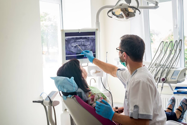
Dental beam CT (computer tomography) is an x-ray
machine that is used in 3D imaging by oral surgeons or dentists, when regular dental x-rays don’t yield sufficient results. This
procedure is not performed routinely because the radiations emitted from its
scanners are more as compared to those of regular x-rays found at the dentist.
The CT scanner has special features that enable it
to display three dimensional (3D) pictures of nerve paths, soft tissues, bones,
and dental structures located at the craniofacial area. The high-quality images
produced allows precise planning for treatment. The images are used to evaluate
illnesses that affect the jaw, sinuses, dentition, facial bony structures, and
the nasal cavity.
Common Uses of the Dental Cone Beam
This technique is widely used in the field of
orthodontics to prepare a treatment program routine for each patient. It can
however, be used in extreme cases that will involve:
- Reconstructive surgery
- Evaluation of nerve canals, the nasal cavity, and the jaws
- Pathology, or the identification of the origin/source of pain
- The placement of teeth implants
- Detection and treatment of jaw tumors
- TMJ (temporomandibular joint disorder) diagnosis
- Tooth orientation
How to Prepare
This examination does not need any special
preparation. All that is required of you is to make sure that you don’t have
any metallic objects like jewelry, hairpins, hearing aids, or eyeglasses on
you during the scan as they tend to affect the imaging process and can lead to
a misdiagnosis.
Dentists in Smithfield, Utah, advise people with
temporary dental works to bring them to the oral surgeon or dentist to have
them examined as well. Expectant mothers are also advised against going for the
procedure.
How the Procedure is Performed?
During the examination, you will be requested to
either lie on the examining table or to sit on the examining chair. This will
depend on the type of scanner to be used. You will be positioned by the oral
surgeon in a position that sets the area to be examined at the center of the
beam. While you remain still, the detector and the x-ray source will make a
360-degree rotation while generating 2D images of the dental structures and the
whole mouth with a resolution of up to 200. The images are then combined
digitally to create 3D images that provide oral surgeons or dentists with information
about the patient’s craniofacial and oral health.
The Benefits
- The CT has the ability to image soft tissue and bones in one scan
- It is accurate, non-invasive, and painless
- Provides additional information than dental x-rays
- No radiation will remain in the body of the patient after the examination

Comments
Post a Comment